Developing an MRI machine unlike any you have ever seen
the team behind the Frugal MRI Scanner aim to bring magnetic resonance imaging
to low-resource healthcare settings by making a small, inexpensive and portable scanner.
Learn more about their quest in this Knowledge File
Magnetic resonance imaging
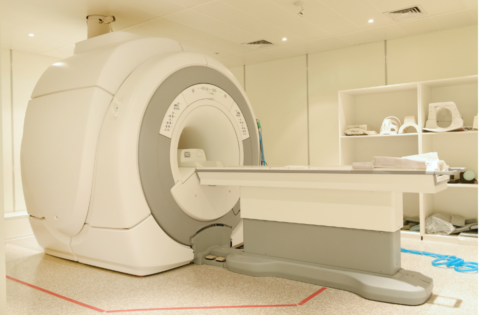
MRI systems, developed in the 1970s, completely transformed medicine. MRI is a non-invasive technique which produces three-dimensional images with exquisite tissue contrast, but there is a huge downside. A typical clinical system needs a very strong magnetic field provided by a superconducting magnet that weighs several tons and needs liquid helium for cooling. The full device can cost up to three million euros, plus one needs an expensive state-of-the-art infrastructure to make it work, and highly-trained technicians to operate them.
When a patient lies in an MRI scanner the water molecules act as tiny magnets, precessing at an atomic level in a similar way to a spinning gyroscope. These precessing magnets create a signal, analogous to the magnet on a bike wheel driving a dynamo, and this signal is picked up by , detectors built into the scanner. By analyzing the frequency characteristics of these signals, three-dimensional image of your insides can be formed.
How to liberate MRI from a heavy and expensive cocoon? Frugalise it.
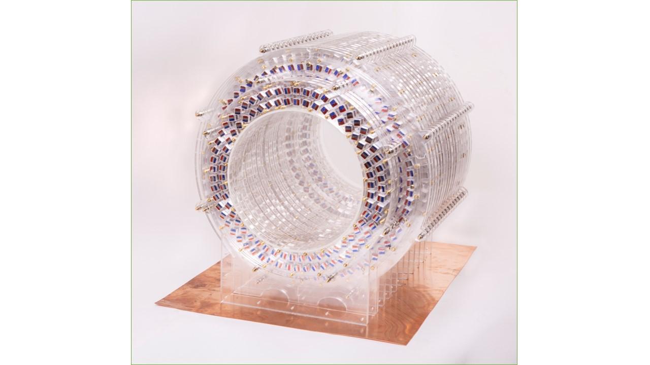
The key step is to take the dominant high cost of the magnet out of the equation. Rather than a superconducting device, design one with small permanent magnets which require no energy and no cooling. This results in a cheaper, lighter and more portable device. Such a low-field MRI scanner opens up applications that you previously would not have thought of. Think about low-resource healthcare settings such as the ambulance, military field hospitals, remote areas, sports arena, or monitoring the treatment of diseases such as hydrocephalus (a serious condition that consists of an accumulation of cerebral fluid that puts pressure on the brain) which are prevalent in developing countries.
When you use a weak magnet it means the received signal is much smaller which means that advanced mathematical methods of reconstructing the images plays a bigger role, and the team from the TU Delft has extensive experience over the past years. The scientists figured out how to build a low-field MRI that makes medically useful pictures by bringing the borders between design, hardware, electronics and advanced mathematics all together. The team worked on several prototypes over the last years and arrived at a prototype that gives images. After improving their quality, both by image processing techniques and hardware improvements, the scanner is now in place.
The multidisciplinary and intercontinental team
The scientists that are involved in the project form a truly interdisciplinary and international team. Applied Mathematician Van Gijzen and Electrical Engineer Remis are from the TU Delft. These Dutch scientist write algorithms behind technical applications and simulations. The British Andrew Webb is Professor in Radiology at the LUMC, Director of the Gorter MRI Center, and is a world specialist on MRI.
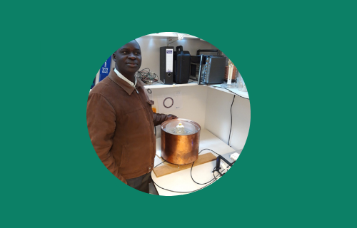
Steven Schiff is head of the Neuroengineering Department of Pennsylvania State University and manages a large research programme on hydrocephalus in Uganda. Johnes Obungoloch is vice-dean of the biomedical engineering department at Mbarara University in Uganda. Over the years, many early career scholars affiliated with the Leiden-Delft-Erasmus universities have been part of the research team. Together they form a very diverse and inspiring group of people.


The goal: extending MRI from the hospital into primary healthcare
The team behind the low-field scanner started to work together with a specific goal: to monitor progress after treatment of paediatric hydrocephalus (waterhoofd) in Uganda. In Uganda and in many other Sub-Saharan countries child mortality rates caused by hydrocephalus are still very high. Due to the very high costs and state of the art infrastructure it needs, regular MRI devices are out-of-reach for many (small) hospitals in Uganda. While the scanner was initially developed to detect hydrocephalus, the design, the magnet and the algorithms can also be used for other diseases.
In time the scanner can be manufactured locally on a worldwide scale. The ultimate goal is to broaden the base for medical science development in both developed and developing countries. In April 2021, Prof. Andrew Webb received 2.4 million euros from the European Research Council. With this Advanced Grant Webb titled PASMAR (Portable and Sustainable Magnetic Resonance) Webb aims to redesign MRI systems for many other applications from the ground-up to increase their accessibility and applicability.
Challenges that require rigorous scrutiny
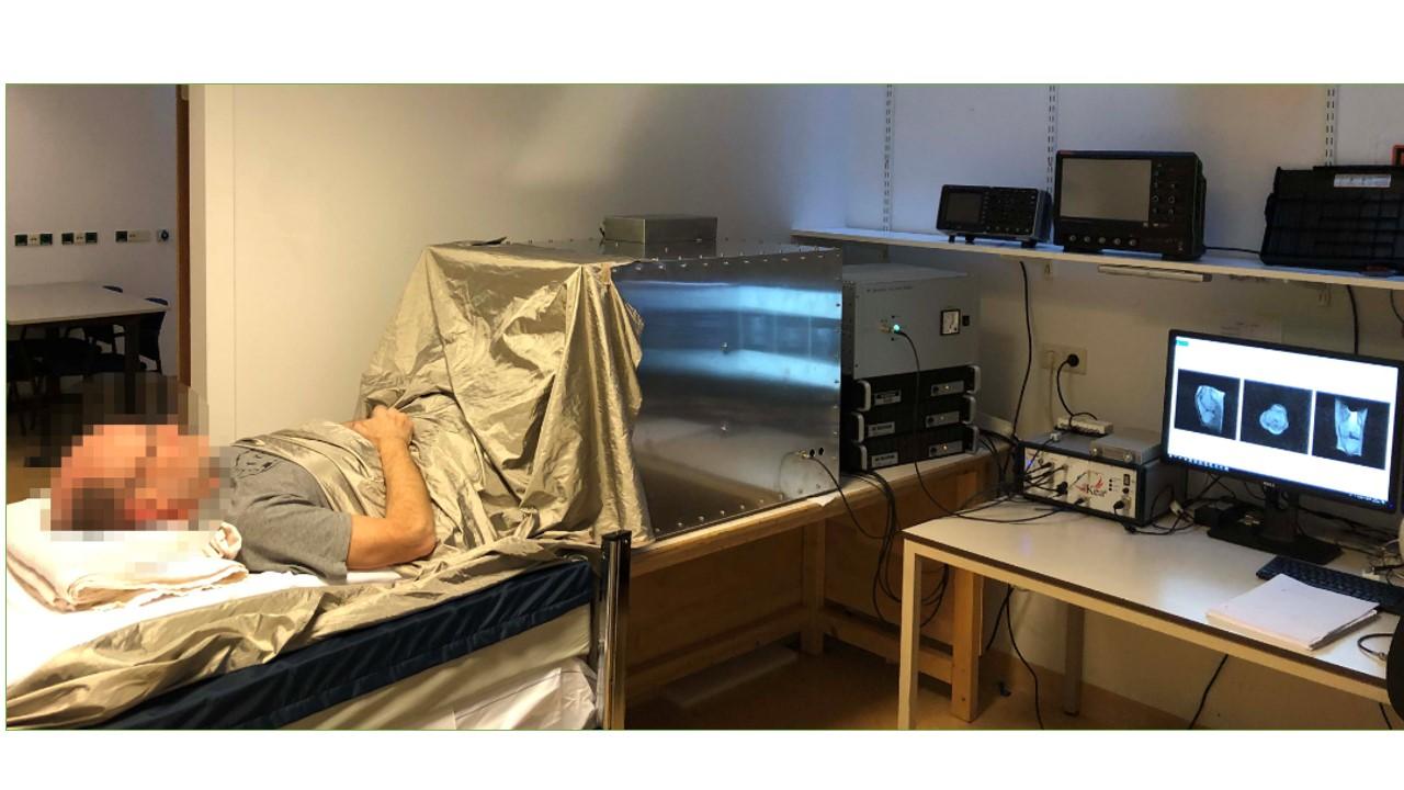
Technically the team has a working prototype that has so far produced high quality images from volunteers. This prototype cost thousands instead of millions of euros to design and construct. The next steps are to perform clinical testing, and amongst other things, to improve the ergonomics and patient-comfort, of the design, as well as ensuring that it can operate within strict clinical guidelines. Many cultural, ethical and entrepreneurial aspects need to be investigated before the low-field scanner can be used in low-resource healthcare settings. There are also many practical issues related to implementation, business models and manufacturing.
The impact on the need of the scanner
A team of early career scholars conducted a qualitative study into the need of such a scanner in healthcare systems in Uganda and patients’ response to such device, and understanding financial and production possibilities in Uganda in order to develop an adequate business model. The study also included the identification of local manufacturing partners, and improvements to the design of the MRI device.
A business plan
A business plan is under development by early career scholars that will help the scanner to go from a scientific innovation to a commercial product. This will allow the new MRI technology to have a widespread impact and help as many people as possible. The technology has the potential to extend the use of MRI from the hospital into smaller primary healthcare facilities. This is due to improved safety (low magnetic field), lower power & infrastructure requirements, and lower cost. The innovation also resonates with themes such as access-to-care, tele-radiology and connectivity/modelling.
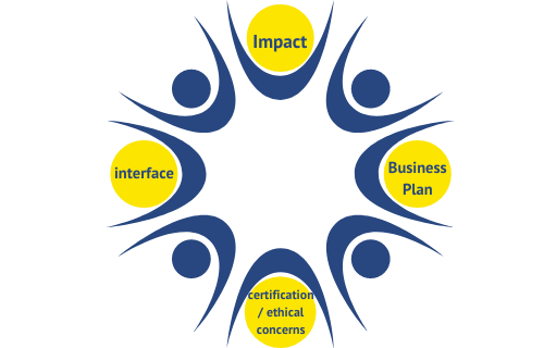
Certification of medical devices and ethical concerns
A team of early career scholars are investigating the certification of medical devices and investigating any ethical concerns. Though these are two different topics, both need to be looked at in order for the scanner to progress towards being tested on patients and ultimately being used in hospitals.
User-friendly interface & open source
A user-friendly web interface that transforms the low the MRI output signals into high quality images is currently in development. The magic algorithms crunching the data have been written by experts at the TU Delft. This website will be key in connecting users and the entire low field programme. As Webb believes it is important that this new development reaches as many people in developing countries as possible, the design of the MRI system will be open to the public. This means the system can also be maintained and repaired locally.